Nov. 14, 2023
UCalgary researchers discover unique differences in cancer cells based on where they are in a tumour

Taking a tumour biopsy is a critical first step used to gather essential information and diagnose disease. Generally, a biopsy is taken from the centre of the suspicious tissue, mass, or lesion; however, a recent University of Calgary study shows cells found at the edge of a tumour should be considered.
“Current technology allows us to look at tumour cells in a new way providing new understanding,” says Dr. Pinaki Bose, PhD ‘10, assistant professor at the Cumming School of Medicine, and principal investigator. “We can see unique differences in cells found in the middle of a tumour compared with those found on the edge, which can help guide treatment.”
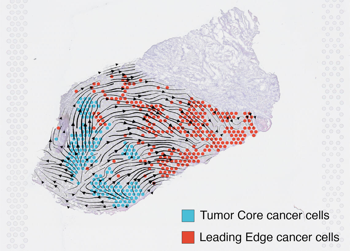
Examining the centre of the tumour is generally the first step in discovery and allows for cancer staging, tumour grading, prognosis, and treatment plan. Using a technology called spatial transcriptomics, a molecular profiling method that preserves the positional identity of cells, the researchers were able to map and see important activity in a tumour including how the organization of cells affects the biology of a cancer. The study, published in Nature Communications, showed important differences in the cells’ architecture.

Pinaki Bose
Analyzing a specific type of mouth cancer, oral squamous cell carcinoma, the researchers found that cells on the tumour’s edge, where it interacts with normal tissue, provided very distinct information compared to cells from the middle. The team believes this precise information could help in providing better understanding of the tumour, its aggressiveness, and the types of therapeutics it could respond best to.
“We also discovered that gene expression patterns in cells on the edge of a tumour aren’t unique to mouth cancers,” says Bose. “Our research showed similar results across several different cancer types, including breast, pancreatic, prostate and lung. This is an unexpected finding and could provide new targets for treatments.”
Findings shows that the gene signature in cells at the edge of the tumour were associated with worse clinical outcomes across most cancer types, while the gene signature in cells at the core were associated with improved prognosis in cancers of similar origin to mouth cancers.

Rohit Arora, left, and Christian Cao.
Bose says the use of spatial transcriptomics data and computational modelling paves the way for a deeper understanding of tumour biology and could unlock new information about how tumours invade healthy cells, what triggers metastasis and how these cells escape the body’s immune response.
The study’s co-first authors, Rohit Arora, BHSc ’23, and Christian Cao were undergraduate students when the study was conducted, and Bose gives them a lot of credit for the important discoveries reported in the study. Both students continue their academic pursuit; Arora is a doctoral student at Harvard University and Cao is a medical student at the University of Toronto.
This research is supported by a Precision Oral Biology (PROBE) grant from the Ohlson Research Initiative and the Precision Oncology and Experimental Therapeutics (POET) Program.

Spatial transcriptomics showed important differences in a tumour cell’s gene expression.
Bose Lab
Pinaki Bose is an assistant professor in the departments of Oncology and Biochemistry & Molecular Biology at the Cumming School of Medicine (CSM). He is a member of the Arnie Charbonneau Cancer Institute, director of tumour biology and translational research at the Ohlson Research Initiative, a research program focused on head and neck cancer at the CSM, and scientific lead of the POET program at the Tom Baker Cancer Centre.


